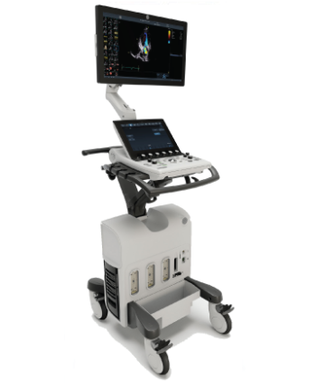
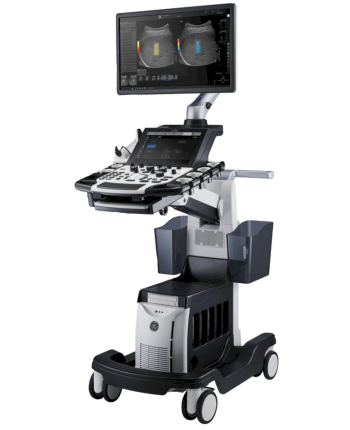
SKU: N/A Categories: , Brand:

GE Voluson P8 Ultrasound Machine


The GE Voluson P8 is an entry-level ultrasound system for women’s healthcare applications. Its automated functions allow physicians in busy practices to maintain high-quality imaging while reducing the time needed to conduct, analyze, and report on exams. The consensus of the GE Voluson P8 is an affordable ultrasound for any medical professional who wants to experience the power of a GE Voluson system for clinical applications. Even though the GE Volsuon P8 system is at a lower price point, it supports in-depth imaging features, such as 3D/4D and HDlive. In refurbished condition, Voluson P8 prices range from about $16,000 to $26,000, depending on the condition, age, iteration (BT15/BT16), degree of use, and what transducers come with the system. Scroll down for more details about GE Voluson P8 features, specifications, and reviews. Contact Ultrasound Supply, a Probo Medical company and a leading seller of refurbished GE Voluson ultrasounds, for more information.

Clear

Share it:

General Product Information - Disclaimer

All items mentioned below including probes, accessories, options, features and similar properties are solely meant to provide general information about the maximum capability of the product. Some or all features mentioned may not be included in the listed price. Please contact us directly for accurate pricing per each additional feature.

Overview

Application Training for the GE Voluson P8

Our on-staff sonographer can provide you with onsite applications training for the GE Voluson P8 at a set price plus travel costs.

Voluson P8 Ultrasound Service Options

Free technical support is available during the installation of the ultrasound system as well as over the course of the standard limited warranty. Additional technical support is available after the warranty period expires at an hourly cost per issue.

GE Voluson P8 Maintenance Recommendations

Ultrasound Supply recommends using a surge protector along with a dedicated power outlet on the Voluson P8. To ensure proper sanitation and patient health, probes should be disinfected after each use with a medical-grade disinfectant wipe that’s safe for ultrasound lenses.

Dimensions & Weight of the GE Voluson P8

Width: 16.5 in (420 mm)
Depth: 626.4 in (70 mm)
Height: 59 in (1495 mm)
Weight: 143 lbs (65 kg)

GE Voluson P8 Specifications

System processing channel: Counts up to 161,616 by configuration
Minimum depth of field: 0 – 1 cm (Zoom, probe dependent)
Maximum depth of field: 0 – 30 cm (probe dependent)
Continuous dynamic receive focus/continuous dynamic receive aperture
Adjustable dynamic range: Up to 255 dB

GE Voluson P8 Electrical Power

Nominal input voltage: 100-240 VAC, frequency 50/60 Hz

Review

The GE Voluson P8 is an entry-point to the Voluson ultrasound family, allowing physicians to enjoy all the technological benefits of GE’s innovations over the decades at an affordable price point of around $16,000 to $26,000. The Voluson P8 was introduced in 2012 and developed based on the Voluson S6, Voluson S8, and the Voluson E-Series, which includes the highly popular Voluson E8 and Voluson E10. The imaging concept and design of the GE Voluson P8 are primarily focused and optimized for OB/GYN applications. They are delivered through GE’s advanced imaging technologies, including Coded Harmonic Imaging with Pulse Inversion technology, Speckle Reduction Imaging, and CrossXBeam (Compound Resolution Imaging). The GE Voluson P8 also provides an intuitive and easy-to-use keyboard from the Voluson family that allows for the immediate performance of routine clinical scanning.

One of the major strengths of the GE Voluson P8 from an efficiency perspective is that it features the integration of several of GE’s unique automation technologies. Auto Optimization for 2D and Doppler helps enhance contrast resolutions at the touch of a button. Additionally, SonoBiometry, SonoNT/IT, SonoAVCfollicle, SonoL&D, and SonoRenderStart all improve the scanning and diagnosis efficiency of key exam operations by reducing the number of keystrokes needed as well as enhancing the reproducibility of measurements.

3D HDlive is one of the primary features that set the GE Voluson P8 apart from many of its competitors, such as the Samsung UGEO H60 and SonoAce R7 and the Philips ClearVue 650. Hospital administrators and specialized clinics will also find this high-resolution 3D imaging technology cost-efficient, with added value for physicians and patients.

Probes

GE Voluson P8 Probes and Transducers

Convex Probes:
4C-RS

Linear Probes:
12L-RS

Endocavitary Probes:
E8C-RS

Phased Array Probes:
3Sc-RS

3D/4D Convex Probes:
RAB2-6-RS

3D/4D Endocavitary Probes:
RIC5-9W-RS

Features

GE Voluson P8 Features

• 17” High Resolution LCD LED Flat Panel Display
• 3 Active Probe Ports
• Integrated HD (500 GB)
• 3 USB Ports for External Peripherals
• 2 USB Ports for On-board Peripherals
• AO(Automatic Optimization)
• CrossXBeam(Spatial Compounding Imaging)
• SRI(Speckle Reduction Imaging)
• B-Steer
• Coded Phased Inversion Harmonic Imaging
• HD-flow
• 3/4D(Real time)
• Virtual Convex
• Patient Information Database
• SonoBiometry
• SonoRender Start
• Raw Data file

Measurement & Calculations (Including Worksheets & Reports)

OB-GYN
Vascular
Cardiac
Abdominal
Small-Parts
Urology
Pediatrics
Musculoskeletal
Neurology

Accessories

Peripheral Options for the GE Voluson P8

• Sony Digital UP-D711 Termal Printer
• Sony Fixture Kit for Digital UP-D711 Thermal Printer
• Sony Digital UP-D25 Color Thermal Printer
• Sony Digital UP-D897 BW Thermal Printer
• Mitsubishi P93W/E Thermal Printer
• Mitsubishi P95DW Thermal Printer
• Footswitch MKF2-MED USB GP26
• USB ECG Kits (AHA/IEC)

Standard Voluson P8 Supplies

• Aquasonic Ultrasound Gel
• Sono Ultrasound Wipes
• Sony UPP-110HG Thermal Printing Paper
• Sony UPC-21L Color Thermal Printing Package
• Mitsubishi KP95HG Thermal Roll Paper
• Mitsubishi KP65HM-CE High-Density Thermal Paper

GE Voluson P8 Ports

• 3 USB Ports for External Peripherals
• 2 USB Ports for On-board Peripherals
• RJ45 LAN Port
• 1 HDMI Out Port
• 1 AUDIO Out Port

GE Voluson P8 Image Storage Formats & Devices

Storage Formats:

– DICOM files (Single- or multi-frame) DCM and DICOM Files with DICOMDIR

– Raw Data File (Proprietary format)

– Export Data as BMP, TIFF, JPEG, MPEG 4, or MS Video 1

Storage Devices: USB Memory Stick
DVD-RW Storage
HDD Image Storage

Options

Options for GE Voluson P8 Ultrasounds

• Static 3D Mode
• 3/4D Advanced
• HD Live(3D Only)
• SonoNT/SonoIT
• SonoL&D
• 4D View PC Software
• DICOM 3
• Extended View(XTD)
• Anatomical M-mode
• Report Editor

Applications

GE Voluson P8 Clinical Applications

Abdominal
Gynecology
Small-Parts
Vascular
Pediatrics
Cardiology
Urology
Musculoskeletal (MSK)
Breast

Technical Vocabulary

Definitions for Key GE Voluson P8 Ultrasound Features

Auto Optimization is a one-touch image optimization function that allows a user to optimize the image based on the actual B-Mode image or Pulse wave Doppler data. This function works based on preset levels (Low, Medium, and High) and allows users to pick a preference for the contrast enhancement in the resulting image. Low level offers the least amount of contrast enhancement, while High does the most. Auto Optimization is available in single or multi image, on live, frozen or CINE images (in B-Mode only), while in zoom, and in Spectral Doppler. Auto Optimization in PW Doppler Mode also optimizes the spectral data. The setting auto adjusts the Velocity Scale (for live imaging only) and offers baseline shift, dynamic range, and invert (if preset). Upon deactivation, the spectrum is still optimized.

SRI (Speckle Reduction Imaging) reduces speckle noise in images. It affects edges and fine details, which limit the contrast resolution and make diagnostic more difficult.

CrossBeam (Spatial Compounding Imaging) obtains real-time sonographic information from several different angles of insonation and combines them to produce a single image. CrossBeam helps reducing speckle artifacts, enhancing mass margin delineation, and improving anatomical details.

Coded Harmonic Imaging (CHI) utilizes Digitally Encoded Ultrasound (DEU) and enhances near-field resolution for improved small-parts imaging. CHI diminish low-frequency, high-amplitude noise, and improves imaging technically on difficult patients. Harmonics may be especially beneficial when imaging isoechoic lesions in shallow-depth anatomy in the breast, liver, and hard-to-visualize fetal anatomy. Harmonics may also improve B-Mode image quality without introducing a contrast agent.

Virtual Convex is available on linear and Sector probes. Virtual Convex provides a larger field of view in the far-field and is always active with sector probes.

Extended View (XTD) enables a transducer to be moved along a larger organ, stitching multiple images together to form one long image with an extremely wide field of view.

Raw Data is a software tool that enables image processing, quick data re-acquisition, and image analysis with the same resolution and same frame rates of original images. Raw Data helps shorten exam duration, improves clinical workflow by post-processing, and reduces the time to put the probe on a patient.

SonoBiometry performs a semi-automatic measurement of the head both head circumference and bi-parietal diameter), abdomen and femur. This tool can help enhance clinical workflow by helping reduce keystrokes to perform biometry measurements.

SonoNT (Sonography-based Nuchal Translucency) and SonoIT† (Sonography-based Intracranial Translucency) are Voluson technologies that help provide semi-automatic, standardized measurements of the nuchal and intracranial translucency as early as 11 weeks. Both tools can integrate easily into your workflow. SonoNT helps avoid the inter- and intra-observer variability that comes with manual measurements and helps provide you with the reproducibility you demand.

SonoAVC*follicle (Sonography-based Automated Volume Count follicle) automatically calculates the number and volume of hypoechoic structures in a volume sweep, helping improve efficiency and workflow of follicular assessment. This feature helps to detect low echogenic objects (eg., follicles) in an organ (eg., ovaries) and analyzes their shape and volume. From the calculated volume of the object, an average diameter will be calculated. All objects detected that way will be listed according to size. The calculation results are displayed in the right monitor area. The objects are listed according to size. All different objects are color-coded, i.e., the color surrounding the number of the object also denotes the object on the image. If the mouse cursor hovers over a specific item on the list the respective object in the image is highlighted and vice versa. The color of the object is bound to its position on the list.

Battery Pack is an optional power supply. The system power is maintained by the battery when there is an AC power failure or the power cable is unplugged. The battery pack also helps to maintain the Voluson P8 ultrasound system’s power when it needs to be run for long durations of imaging.

Year Launched

2012

Estimated Market Price

Economy

Monitor

17" LCD

Tilt/Rotate Adjustable Monitor

Tilt/Rotate only

Monitor Resolution

1280*1024

Image Size Resolution

800

Touch Screen

No

Trackball or Trackpad

Trackball

CP Back-Lighting


Weight

143lbs (65kg)

Probe Ports

3

Battery

Yes

Boot-Up Time


Sleep Mode

Yes

Maximum Depth of Field

30cm

Minimum Depth of Field

0-1cm

Cart

No

Independent Steer & Lockable Wheels

Yes

2D, M mode

Yes

M-color Flow Mode

Yes

Anatomical M-mode

Yes

Trapezoidal Mode

Yes

Color, Power Angio, Pulse Wave Doppler

Yes

Bi-directional Power

Yes

SCW Doppler

Yes

Tissue Doppler(Velocity) Imaging

No

Freehand 3D

Yes

Live 3/4D OB/GYN

Yes

HD Live

Yes

STIC

No

Live 3D Echo

No

Stress Echo

No

Strain and Strain Rate

No

B Flow

Yes

Panoramic Imaging

Yes (XTD)

Contrast Imaging-Cardiac

No

Contrast Imaging - General Imaging

No

Strain-based Elastography

No

Shear Wave Elastography

No

Tissue Harmonic Imaging

Yes

Spatial Compounding(=CrossXbeam)

Yes

Speckle Reduction (=SRI)

Yes

Auto Image Opt(B mode)

Yes

Auto Image Opt(Doppler)

No

Write Zoom

Yes

Triplex Mode

Yes

Needle Enhancement or Needle Recognition

No

Auto NT Measurement

Yes

Auto Follicle 2D Measurement

Yes

Auto Follicle 3D Measurement

Yes

Auto IMT

No

Auto IMT (Real Time)

No

Automated B/M/D Measurement

No

Automated LH Measurement(Automated Function Imaging(AFI), Cardiac Motion Quantification(CMQ), or Auto EF(Ejection Fraction)

No

Live Dual (B/BC) Mode

No

SmartExam or Scan Assistant

No

Fusion

No

Raw Data File

Yes

Flexible Report

Yes

Barcode Reader

No

Gel Warmer

No

Abdominal

Yes

Women's Health Care

Yes

OB

Yes

Fetal Echo

Yes

Vascular

Yes

TCD(Transcranial)

No

Small Parts

Yes

MSK/Anesthesiology

Yes

Pediatrics

No

Urology

No

Echocardiography_Adult

Yes

Interventional Cardiology

No

Echocardiography_Pediatric

No

Echocardiography_Neonate

No

Stress Echocardiography

No

Transesophageal Echo_Adult

No

Transesophageal Echo_Pediatric

No

Internal Medicine w/ Shared Service

Yes

Surgury

No

Interventional Radiology

No

Contrast Imaging_General Imaging

No

Contrast Imaging_Cardiac

No

Bowel Imaging

No

Strain Elastography

No

Shear Wave Elastography

No

Convex (1~6Mhz)

Yes(4C-RS)

Convex (2~9Mhz)

No

Single Crystal Convex (1~6Mhz)

No

Single Crystal Convex (2~9Mhz)

No

Single Crystal Micro Convex

No

2D Arrary 3D Convex

No

Micro Convex (5~8Mhz)

No

Single Crystal Endocavity_Straight Type

No

Endocavity_Curved Type

Yes (E8C-RS)

BiPlane Endo Rectal

No

Biplane Micro Convex (3~10MHz)


3D Convex

Yes (RAB2-6-RS)

3D Convex Light Weight

No

3D Endocavity

Yes (RIC5-9W-RS)

3D Micro Convex

No

3D Linear

No

Linear (>14Mhz)

No

Linear (3~12Mhz)

Yes (12L-RS)

Linear (<9Mhz)

No

Single Crystal Linear

No

Single Crystal Linear (3~12Mhz)

No

Single Crystal Linear (<9Mhz)

No

Linear 50mm

No

Linear 25mm

No

Hockey stick (<13Mhz)

No

Hockey stick (>13Mhz)

No

T or L shape Intra Operative

No

Laparoscopic

No

Phased Array_Adult

Yes (3Ss-RS)

Single Crystal Phased Array_Adult

No

2D Arrary 3D Phased Array

No

Phased Array_Pediatric

No

Single Crystal Phased Array_Pediatric

No

Phased Array_Neonate

No

ICE

No

TEE_Adult

No

TEE_Pediatric

No

2D Array 3D TEE

No

Pencil CW (2Mhz)

No

Pencil CW (5 or 6Mhz)

No

DICOM 3.0

Yes(Option)

DICOM SR_Cardiac

Yes

DICOM SR_Vascular

Yes

DICOM SR_OB/GYN

Yes

JPEG, WMV, & AVI

Yes

USB

Yes

HDD/SDD

500GB

DVD/CD RW

Yes

Wireless LAN

No

GE Voluson P8 Ultrasound Review Video

Watch a short video review of the Voluson P8 ultrasound system by our parent company, Probo Medical.