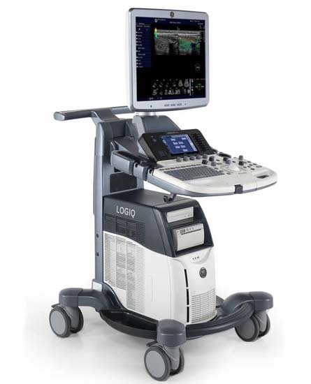
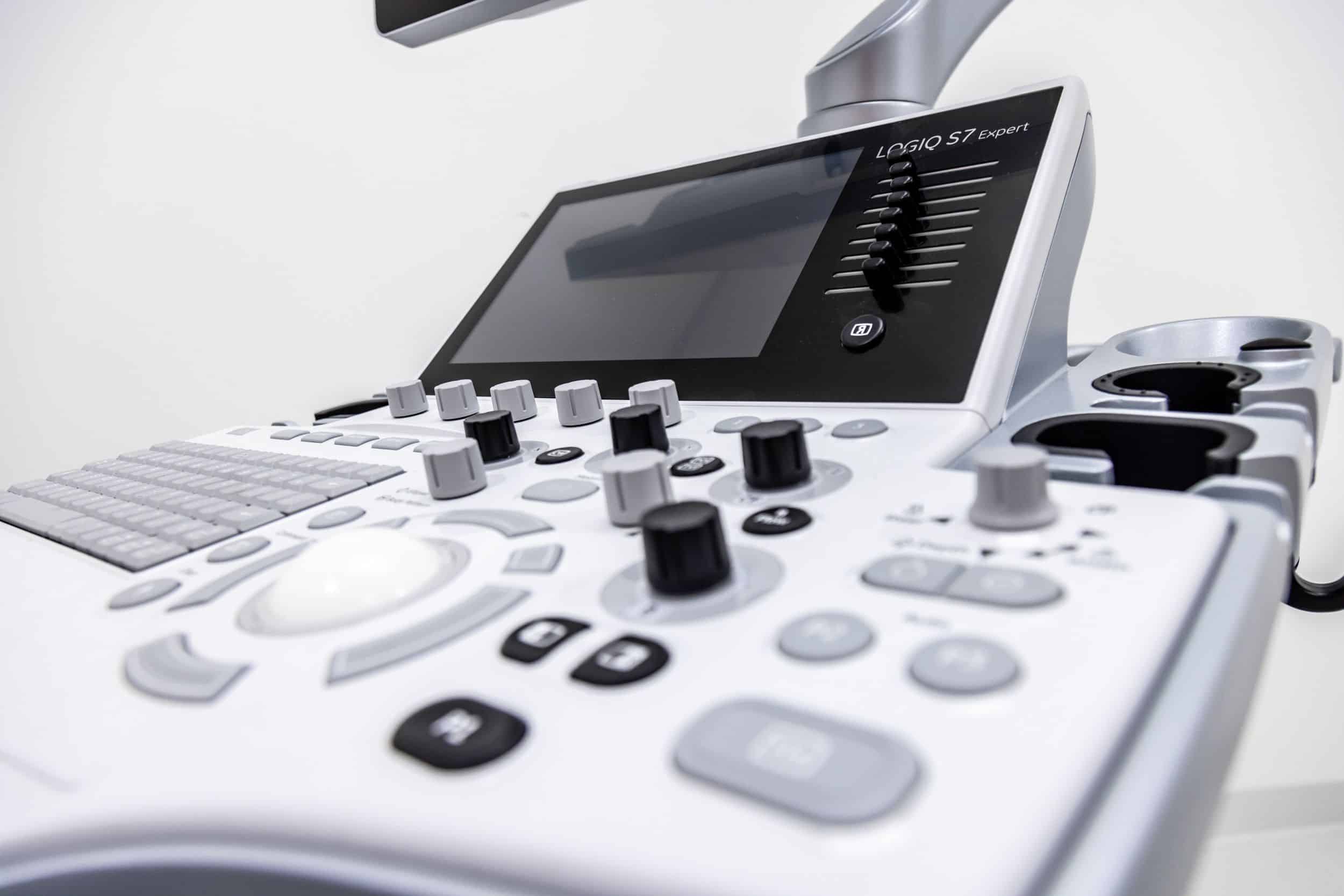
GE Logiq S7 With XDClear Dimensions and Weight:
• Height: 1,210mm (Minimum), 1,760mm (Max.)
• Width: 530mm (Caster), 520mm (Keyboard), 565mm (Monitor)
• Depth: 865mm (Max.), 795mm (Caster)
• Weight: 90 kg (198 lb.)
GE Logiq S7 With XDClear Specifications:
• Digital beamformer
• Displayed Imaging Depth: 0 – 33 cm
• Minimum Depth of Field: 0 – 2 cm (Zoom) (probe dependent)
• Maximum Depth of Field: 0 – 33 cm (probe dependent)
• Continuous dynamic receive focus/continuous dynamic receive aperture
GE Logiq S7 With XDClear Electrical power:
• Voltage:100-120 Vac or 220-240 Vac
• Frequency: 50/60 Hz
• Power: Consumption maximum of 900 VA with peripherals
GE Logiq S7 XDClear Review:
The GE Logiq S7 with XDclear, as with the GE Logiq S8 with XDClearwith XDclear, was developed on S-Agile Architecture, one of GE’s most advanced ultrasound platform. Besides the S7 having a larger 10.1” LCD touch screen, the two systems are identical architecturally. With revision 3.0, the accuracy and reliability of the S7 can reach a whole new level, thanks to GE’s single crystal XDclear transducers such as the C1-6-D and C3-10-D. Ultrasound operators can enjoy premium image resolution, penetration, and wide bandwidths that were only supported by the GE Logiq E9 before.
If you’re looking for a system with a great platform, but don’t need premium options or dedicated transducers, then the GE Logiq S7 with XDclear is an excellent choice. To fully benefit from its superb cardiac functionality, the scan Assistant, Auto EF, and Cardiac Automated Function Imaging (cardiac AFI) options can be added to this shared service system. As with the GE Logiq S8 with XDclear, the price point of the GE Logiq S7 with XDclear is still a bit high compared to similar system such as the Philips Affiniti 50. The decision to pick the right system can be even more difficult if you have a manufacturer refurbished Philips HD11XE or HD15 system as an option.
GE Logiq S7 XDClear Probes/Transducers:
XDClear Probes
C1-6-D [1-6 MHz]
C3-10-D [2-11 MHz]
Convex Probe
C1-5-D [1-6MHz]
Intra-Cavitary Probe
IC5-9-D [3-10MHz]
Micro-Convex Probe
3CRF-D [2-5MHz]
8C-D [4-11MHz]
10C-D [4-12MHz]
Linear Probe
9L-D [2-8MHz]
11L-D [3-11MHz]
L3-12-D [2-11MHz]
ML6-15-D [4-15MHz]
L8-18i-D [4-15MHz]
Sector
3Sp-D [1-5MHz]
S2-5-D [-5MHz]
6S-D [2-8MHz]
S4-10-D [3-9MHz]
Real-time 4D
RAB4-8-D [4-8MHz]
RIC5-90D [3-10MHz]
Speciality
BE9Cs-D [3-12MHz]
6Tc-RS [2-8MHz]
P2D [2MHz]
P6D [6.3MHz]
P8D [8.3MHz]
GE Logiq S7 XDClear Features:
• High resolution 23″ wide LCD monitor
• High resolution 10.1″ wide LCD touch screen
• 4 active probe ports and 1 parking probe port
• Integrated HDD
• Integrated DVD Multi Drive
• Integrated speakers
• Locking mechanism that provides rolling lock and caster swivel lock
• Integrated cable management
• Front and rear handles
• Easily removable air filters
• Automatic Optimization
• CrossXBeam
• Speckle Reduction Imaging (SRI-HD)
• Fine Angle Steer
• Coded Harmonic Imaging
• Virtual Convex
• Advanced 3D
• Patient information database
• Image Archive on integrated CD/DVD and hard disk drive
• Raw Data Analysis
• Real-time automatic Doppler calcs
• OB Calcs
• Fetal Trending
• Multi gestational Calcs
• Hip Dysplasia Calcs
• Gynecological Calcs
• Vascular Calcs
• Urological Calcs
• Renal Calcs
• Cardiac Calcs
• inSite ExC Capability
• On-board electronic documentation
• MPEGVue
• Key Macro
• Network Storage
• Quick Save
• Quick Patient Entry
• Email2MMS
GE Logiq S7 With XDClear Peripheral Options:
• Sony Digital UP-D711 Termal Printer
• Sony Fixture Kit for Digital UP-D711 Thermal Printer
• Sony Digital UP-D25 Color Thermal Printer
• Sony Digital UP-D897 BW Thermal Printer
• Mitsubishi P93W/E Thermal Printer
• Mitsubishi P95DW Thermal Printer
• Printer installation kit
• External USB printer connection
• Printing using Bluetooth® connectivity (Inkjet printer option)
• Wireless LAN
• HDMI output available for compatible devices
• Footswitch with programmable functionality
• Universal Video Converter
• Power Assistant
• Battery Pack (for Power Assistant)
• Isolation Transformer
• Isolated 1Gb Ethernet connection
• Isolated USB connector
• EMI filter (PWR supply noise filter)
• RS-DLP Probe Adapter for TEE Probe
• Side Cabinet (Japan only)
• Drawer
• Small Probe Holder (Probe Holder Adapter for Small Probe)
• Multi Purpose Holder (Vertical Endocavitary Probe Holder)
• Optional Probe Holder (Side Probe Holder)
• Probe cable hanger
• ECG + AHA/IEC® Cables
• USB 3 Pedal Foot Switch
GE Logiq S7 With XDClear Supplies:
Aquasonic ultrasound gel
Sono ultrasound wipes
Sony UPP-110HG thermal printing paper
Sony UPC-21L color thermal printing pack
Mitsubishi KP95HG thermal roll paper (new)
Mitsubishi KP65HM-CE High density thermal paper
GE Logiq S7 With XDClear ports:
• User accessible USB x 5 ports
• HDMI connector
• Ethernet 1000/100/10BaseT
• Audio Line Out (1.5mm pin jack)
GE Logiq S7 With XDClear image storage:
• On-board database of patient information from past exams
• Storage Formats:
• DICOM – compressed/ uncompressed, single/ multiframe, with/ without Raw Data
• Export JPEG, JPEG2000, WMV (MPEG 4), and AVI formats
• Storage Devices:
– USB Flash Device: 64MB to 64GB (for exporting individual images/clips)
– CD-R storage: 700MB
– DVD storage: R (4.7GB)
– Hard Drive Image Storage: ~300GB
– Compare previous exam images with current exam images
– Reload of archived data sets
– Network Storage support for Import, Export, DICOM Read, SaveAs, SaveAs
GE Logiq S7 with XDClear Applications:
• Fetal/Obstetrics
• Abdominal (includes renal, GYN/Pelvic)
• Pediatric
• Small Organ (breast, testes, thyroid)
• Neonatal Cephalic
• Adult Cephalic
• Cardiac (adult and pediatric)
• Peripheral Vascular
• Musculo-skeletal Conventional and Superficial
• Urology (including prostate)
• Transrectal
• Transvaginal
• Transesophageal
GE Logiq S7 with XDClear FAQs:
• Auto Optimization is a one-touch image optimization function that a user uses to optimize the image, based on the actual B-mode image or Pulse wave Doppler data. The function works based on preset levels (Low, Medium, and High) and allows users to pick a preference for the contrast enhancement in the resulting image. Low does the least amount of contrast enhancement, high does the most. Auto Optimization is available in single or multi image, on live, frozen or CINE images (in B-Mode only), while in zoom, and in Spectral Doppler. Auto Optimization in PW Doppler Mode optimizes the spectral data. Auto adjusts the Velocity Scale (live imaging only), baseline shift, dynamic range, and invert (if preset). Upon deactivation, the spectrum is still optimized.
• SRI (Speckle Reduction Imaging) reduces speckle noise in the images, which affect the edges and fine details that limit the contrast resolution and make the diagnostic more difficult.
• CrossBeam (Spatial Compounding Imaging) obtains real time sonographic information from several different angles of insonation, and combines them to produce a single image. CrossBeam helps reduce speckle artifacts, enhance mass margin delineation, and improve anatomical details.
• Coded Harmonic Imaging (CHI) utilizes Digitally Encoded Ultrasound (DEU). Harmonics enhances near field resolution for improved small parts imaging. Harmonics diminishes low frequency high amplitude noise and improves imaging for technically difficult patients. Harmonics may be especially beneficial when imaging isoechoic lesions in shallow-depth anatomy in the breast, liver, and hard-to-visualize fetal anatomy. Harmonics may improve B-Mode image quality without introducing a contrast agent.
• Virtual Convex is available on linear and Sector probes. Virtual Convex provides a larger field of view in the far field. Virtual Convex is always active with sector probes.
• Raw Data is a software tool that enables image processing, quick data re-acquisition, and image analysis with same resolution and same frame rates of original images. Raw Data shortens exam duration, improves clinical workflow by post-processing, and reduces a time to put the probe on a patient.
• Battery Pack is an optional power supply. The battery maintains the system’s power when there is an AC power failure or when the power cable is unplugged. Also, this helps maintain the system’s power when it needs to be.
• LogiqView (Panoramic Imaging) enables a transducer to be moved along a larger organ, stitching multiple images together to form one long image with an extremely wide field of view.
• Auto IMT automates measurement of intima-media thickness of vessels. Auto IMT helps keep track of atherosclerosis diseases from the early stage as it is developed.
• B Steer simply slants B-mode or Color Flow linear images left or right to get more information without moving the probe. This enables enhanced needle visualization in real time for needle guidance procedures. The angle steer function only applies to linear probes.
• B-Flow utilizes gray scale imaging to visualize a blood flow with different gray intensities according to the reflectors speed and hemodynamics. B-Flow is less dependent on the user or scanning angle, while Color Doppler is heavily dependent on the scanning angle. It also provides higher frame rate and spatial resolution than Color Flow. B-Flow may help visualize vessel-wall irregularities, kidney perfusion, liver and spleen vasculature, and bladder reflux or jets.
• STIC (Spatio – Temporal Image Correlation) imaging presents the heart beating in a multi-planar display while also preserving spatial and temporal relationships of fetal heart. STIC imaging supports captures in grayscale, 3D color, CPA, and directional CPA. The user configures acquisition time, and the function is available on V6-2 volume transducer.
• Anatomical M-Mode helps improve accuracy of arrhythmia assessments and cardiac measurements. It enables to provide M Mode images truly perpendicular to ventricular septum.
• Auto EF is the function that automatically measures LV ejection fraction. Along with the Automated Function Imaging (AFI) feature, Auto EF maximizes time efficiency by the reproducible quantitative output that increases the physician’s diagnostic confidence.
• Automated Function Imaging (AFI) is a decision support tool for regional and global assessment of the LV systolic function. AFI is a tool derived from 2D Strain, which calculates the myocardial tissue deformation based on feature tracking on 2D grey scale loops.
• Scan Assistant is the customable scanning assistant tool that enables a user to save scanning protocols of each lab that helps decrease key stokes from up to 60~70% of ordinary operations. With fewer user’s actions, an exam can be complete because a user does not need to move all around the console, and only requires one button to be pushed at most of the exams.
• Compare Assistant views past studies and current images together in real time via a split screen on the monitor.
• Strain Elastography is a non-invasive diagnostic technique that displays the relative elasticity of tissue stiffness compared to the surrounding tissue by a real-time color map that is superimposed on conventional gray scale image. Most malignant lesions have a harder or stiffer consistency than the surrounding benign tissue. This change in stiffness can also be present in chronic or inflammatory diseases. To displace the underlying anatomical structures, Strain Elastography requires manual palpation by the user, or compression or decompression of the target produced by the patient respiration. It can be an efficient tool to improve biopsy targeting and imaging, evaluate breast and testicular lesions, and perform a quick monitoring and follow up of interventional procedures. It can also provide additional information to increase diagnostic confidence for musculoskeletal diseases such as tendinopathies, tendinosis, synovial hypertrophy, or tears.