Application training
Our on-staff sonographer can provide onsite applications training at a set price plus travel costs.
Service for the Philips iE33
Free technical support is available during installation and over the course of the standard limited warranty. Technical support is available after the warranty period at an hourly cost per issue.
Maintenance Recommendations
Ultrasound Supply recommends the use of a surge protector along with a dedicated power outlet. Probes should be disinfected after every use with a disinfectant wipe proven not to damage the lens.
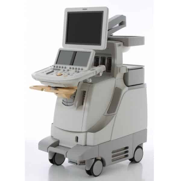
Philips iE33 Dimensions & Weight
Height: (adjustable, maximum) 1626 mm (64 in), (minimum) 1397 mm (55 in)
Width: 559 mm (22.0 in)
Depth: 1092 mm (43 in)
Weight: (no Peripherals) 150 kg (331 lbs.), approx. 480 lbs with packaging
Specifications
Philips xSTREAM imageformer architecture
Up to 442,176 total digital channels (xMATRIX configuration), 57,000 total digital channels (non xMATRIX configuration)
Displayed Imaging Depth: 1 – 39 cm (transducer dependent)
Up to 180 dB system dynamic range
Electrical Power
Voltage 100V-127V, 220V-240V
Frequency 50/60 Hz
Power consumption: 750VA – 900VA depending on system configuration
Philips iE33 revisions: A cart – C cart
Philips first launched the iE33 in 2004 as their new premium shared service ultrasound machine, replacing older units like the Sonos7500. This first version of hardware was designated an “A-cart.” This first version had dual motherboards and dual power supplies and a huge amount of problems with both. The iE33 B-cart improvements were to circuit boards, cables, power supply assembly, and USB ports. The iE33 C-cart brought a new 20” LCD monitor and articulating arm as well as new circuit boards. The R-cart is a refurbished A, B, or C-cart with hardware and software upgrades to G4.0 software.
Philips iE33: D cart – F cart
The iE33 D-cart was launched in 2007 and finally fixed the problems with the dual motherboards by using a unified motherboard. D.1 and D.2 included minor hardware updates. The iE33 E-cart made upgrades to the cart, wheels and locking mechanism. E.1 was a minor hardware & software update. E.2 added major power supply improvements to support Matrix transducers. E.3 was a minor update to the way the iE33 was manufactured. The iE33 F-cart was a major upgrade that added Type III channel boards. F.1 was a minor manufacturing update. F.2 upgraded the video card. F.3 upgraded to a new SATA DVD drive.
Philips iE33: G cart
The iE33 G-cart was launched in 2010 and is the latest version of the iE33. This revision added a new channel board and a 21” widescreen monitor. The iE33 G.1 revised the control panel and added larger touch screens. Ultrasound Supply recommends buying D-cart and up as most hardware problems with the iE33 were fixed by this revision and D-carts can be updated to the latest software
Philips iE33 Vision software updates
The software of an iE33 can be updated to a point limited by the hardware cart. Each major revision of the software was designated with the year and “Vision” in the name. “Vision 2012” was the last major release of the software. Very early A-C cart had software that was named simply “system update” with a number. System update 1.0-2.0 have almost all been upgraded over the years and system 3.0 is the lowest version still commonly found on used iE33s.
The Philips iE33 Vision 2007 software update included support for the new L15-7io intraoperative linear transducer, the X7-2 xMatrix pediatric cardiac, and C8-5 microconvex. Tissue specific presets were added to the S5-1 Purewave cardiac sector probe, and the S12-4 and S8-3 were optimized for 2D and Doppler performance. Improvements were made to LVO and QLAB. Features added were TCD (transcranial Doppler) imaging, Fetal Echo Analysis and Live 3D (4D) Echo.
The Philips iE33 Vision 2008 software update included support the new Purewave C5-1 convex transducer. Features added were TMQ (tissue motion quantification) in QLAB, Viewforum stand alone image viewing platform, improved needle visualization, and contrast capabilities.
The Philips iE33 Vision 2009 software update included support for the new S8-3t pediatric micro TEE probe which is 1/3 the size of the S7-2t. Features added were tissue aberration correction, contrast enhanced ultrasound, and SmartExam automation software.
The Vision 2010 software update on the Philips iE33 added xMatrix including support for the new Purewave xMatrix X5-1 transducer, Live xPlane imaging, and 3D Stress Echo, and support for any PACS. (R-carts can have this update, but cannot support the X5-1 transducer.) This iE33 revision also added cardiac diagnostic capabilities related to ischemic disease detection, structural heart disease assessment, as well as systolic and diastolic heart failure and cardiac arrhythmia
The Philips iE33 Vision 2011 software upgraded QLAB with CMQ-Stress replacing TMQ, and with the additions of the Auto Volume tool, GI 3DQ trending, iCrop, and Elevation compounding. Options with Vision 2011 include the xMatrix performance bundle, and Live 3D. (R-carts can have this update.)
The Vision 2012 software on the Philips iE33 adds support for Auto Doppler, Fetal Heart Navigator, and Vascular Plaque Quantification (VPQ).
Revisions of the Philips iE33
Philips iE33 (A.0) with system update 3.0 – Vision 2010 software
Philips iE33 (B.0) with system update 3.0 – Vision 2010 software
Philips iE33 (C.0) with system update 3.0 – Vision 2010 software
Philips iE33 (R.0) with Vision 2007-Vision 2011 software (limited)
Philips iE33 (D.0) with Vision 2007 – Vision 2012 software
Philips iE33 (D.1) with Vision 2007 – Vision 2012 software
Philips iE33 (D.2) with Vision 2007 – Vision 2012 software
Philips iE33 (E.0) with Vision 2007 – Vision 2012 software
Philips iE33 (E.1) with Vision 2007 – Vision 2012 software
Philips iE33 (E.2) with Vision 2007 – Vision 2012 software
Philips iE33 (E.3) with Vision 2007 – Vision 2012 software
Philips iE33 (F.0) with Vision 2007 – Vision 2012 software
Philips iE33 (F.1) with Vision 2007 – Vision 2012 software
Philips iE33 (F.2) with Vision 2007 – Vision 2012 software
Philips iE33 (F.3) with Vision 2007 – Vision 2012 software
Philips iE33 (G.0) with Vision 2011 or Vision 2012 software
Philips iE33 (G.1) with Vision 2011 or Vision 2012 software
Philips iE33 probes / transducers
xMATRIX X3-1 [ 1 – 3 MHz ] 2,400 elements
xMATRIX X5-1 [ 1 – 5 MHz ] 3,040 elements, PureWave
Pediatric xMATRIX X7-2 [ 2 – 7 MHz ] 2,500 elements, PureWave
xMATRIX TEE transesophegeal X 7-2t [ 2 – 7 MHz ] 2,500 elements, PureWave
TEE transesophegeal S7-2 Omni [ 2 – 7 MHz ] 65 elements, 8.64mm
Pediatric TEE transesophegeal S7-3t [ 3 – 7 MHz ] 48 elements, 7.63mm,
Pediatric TEE transesophegeal S8-3t [ 3 – 8 MHz ] 32 elements, 5.2mm shaft diagmeter, multiplane microTEE
ViewFlex Xtra catheter [ 4.5 – 8.5 MHz ] 64 elements, 120° field of view (for ICE)
PureWave cardiac sector S5-1 [ 1 – 5 MHz ] 80 elements, 20.3mm, adult cardiac sector
Pediatric cardiac sector S8-3 [ 3 – 8 MHz ] 96 elements, 15.4mm
Neonatal cardiac sector S12-4 [ 4 – 12 MHz ] 96 elements, 9.78mm
Intraoperative Linear L15-7io [ 7 – 15 MHz ] 128 elements, 23mm, perioperative
Linear L11-3 [ 3 – 11 MHz ] 288 elements, 39mm, vascular applications
Linear L9-3 [ 3 – 9 MHz ] 160 elements, 38mm, vascular applications
Linear L8-4 [ 4 – 8 MHz ] 128 elements, vascular applications
PureWave Convex C5-1 [ 1 – 5 MHz ] 160 elements, 96° field of view, Coded beamforming
Microconvex C8-5 [ 5 – 8 MHz ] 128 elements, 120° field of view, cerebrovascular applications
Microconvex C9-4 [ 4 – 9 MHz ] 192 elements, 108° field of view, abdominal
Pedoff (CW Transducer) D5cwc [ 5 MHz ] Deep venous and arterial applications, non-imaging
Pedoff (CW Transducer) D2cwc [ 2 MHz ] Adult cardiology applications, non-imaging
Pedoff (PW Transducer) D2tcd [ 2 MHz ] Transcranial Doppler applications, non-imaging
Pulse TY-306 [ 0.04 – 300 MHz ] Heartsound Pulse Contact Sensor, Cardiac and vascular
Advanced Philips iE33 transducers: Matrix Pediatric
The Philips iE33 not only offers six PureWave single crystal transducers that includes four xMatrix probes. xMatrix not only improves image quality and penetration since xMatrix is developed based on PureWave technology, but also allows for real-time scanning of multiple planes and cardiac 4D without needing a typical mechanical 4D probe. This speeds the acquisition of 4D cardiac data over a 4D mechanical probe for mainly OB/GYN. C-plane resolution is also improved over a typical 4D mechanical probe. The [ 1 – 5 MHz ] X5-1 transducer for the iE33 represents the apex of xMatrix design. The [ 1 – 3 MHz ] X3-1 and [ 2 – 7 MHz ] X7-2 are early xMatrix probes, while the [ 2 – 7 MHz ] X 7-2t is a TEE probe with xMatrix technology and represents the pinnacle of transesephegeal technology. In addition, the iE33 supports the largest number of pediatric cardiac transducers including the [ 3 – 8 MHz ] S8-3t multiplane micro TEE and four other pediatric cardiac transducers. The ViewFlex Xtra catheter developed by St. Jude Medical is also compatible with the iE33 and is used for ICE where the catheter is inserted inside the heart through the left femoral vein.
Popular Philips iE33 transducers
The most popular cardiac sector transducer for the Philips iE33 is the [ 1 – 5 MHz ] S5-1, a PureWave transducer offering better than normal image clarity even at deep penetration. The amazing X 7-2t is the most popular transesphegeal probe for the iE33 with it’s xMatrix 4D and multiplanar capabilities.
How the Philips iE33 compares with other Philips systems
The Philips iE33 was launched in 2004 as the replacement to the Sonos 7500 and continued in production until 2015. The iE33 is a premium level ultrasound machine that was top of the line till the introduction of the Epiq line in 2013. The Epiq 7 replaced the iE33 and has a larger monitor and larger touchscreen. It also is smaller and lighter than the iE33. The HD15 sits below the iE33 in features, image quality and price and is a shared service ultrasound machine whereas the iE33 is focused only on cardiovascular applications.
Philips iE33 vs iU22
When Philips launched the iE33 it launched the iU22 at the same time and ever since, many medical professionals have wondered what the difference is, and which is best for their needs. The iE33 is focused only on cardio vascular applications where the iU22 is shared service and can be used for women’s health, general imaging as well as cardiac and vascular applications. Because of this the iU22 supports 4D transducers, and several endocaviary transducers, but no neonatal sector or pediatric TEE. The Philips iE33 and iU22 even look nearly identical; with the major distinguishing feature being dual touchscreens for navigation on the iE33 while the iU22 has only one touchscreen.
Other brands competing with the Philips iE33
The Philips iE33 is the premium cardiovascular-focused ultrasound machine of the past decade. Its closest competitor from GE would be the Vivid E9 and both systems are very similar. The iE33 has two touchscreens and a larger monitor compared to the Vivid E9 and also supports ICE in the latest release where the Vivid E9 has no ICE probe or support. The closest competitor from Siemens is the SC2000 which offers similar image quality and features but is stronger in ICE while the iE33 has better xMtrix TEE imaging.
The Philips Epiq 7 is the top-of-the-line shared service ultrasound machine from Philips. It offers xMatrix and Purewave single crystal probes, a compact and lightweight design and superior ergonomics and workflow. The Epiq 7 replaces the older Philips iE33 but is shared service rather than only cardiovascular. All the Epiq and Affiniti ultrasound machines look visually very similar and use the same monitor and huge tablet like touchscreen for navigation.
The Acuson SC2000 is the top of the line cardiac ultrasound machine in the Siemens lineup with superior 2D, Doppler quality and unique cardiac 4D capbilities in the market.
The Philips iU22 is a premium shared service ultrasound machine that offers superior image quality through xMatrix transducers that offer the best aspects of single-crystal and 4D transducers. In clinical settings, the Philips iU22 offers top-of-the-line image quality and features available for radiology, OB/GYN, cardiac, vascular, and specialty imaging applications, such as MSK, pediatric, and elastography. (For cardiovascular-specific applications, the nearly identical Philips iE33 is slightly better equipped.) The average price for a used Philips iU22 ultrasound is between $8,100 and $14,100, depending on the year of the system, its condition and degree of past use, and whether transducers are included in the package. While Philips has since replaced the iU22 with the more modern Philips Epiq 5 ultrasound, the price differential between the Epiq 5 and iU22 means the latter continues to be a popular choice for physicians who need high image quality at an affordable price. In fact, the iU22, which was launched in 2004, has a long history of improvements and revisions that have made it the gold standard for over a decade.
The GE Vivid E9 is the top-of-the-line premium cardiovascular ultrasound machine from GE. It has amazing image quality and the latest features including xMatrix and 4D cardiac. The Vivid E9 is the replacement for the popular and well respected Vivid 7. The average cost of a refurbished GE Vivid E9 ultrasound is between $18,800 and $28,800, depending on the condition and past use. Still have questions? More information can be found in this article.
The Philips HD15 is a high-end ultrasound machine for shared service applications with the amazing Purewave single crystal probes. Reliable and high performance for nearly a decade the HD15 is still sought after even after production ended in 2015.
Philips iE33 Features
20” Widescreen Flat Panel LCD display monitor on four-way articulating arm
State-of-the-art ergonomic design
Four transducer ports
Two interactive graphical touch screens
Fully adjustable control panel
xMatrix transducer technology
2D Grayscale imaging
XRES adaptive image processing
Advanced XRES adaptive image processing
SonoCT real-time compound imaging
Auto Doppler
iSCAN intelligent optimization
iFOCUS intelligent focusing technology
iOPTIMIZE intelligent optimization
Cineloop review: real-time and duplex modes of up to 2,200 frames
QuickSAVE feature
Exam management features
On-board patient exam storage: Combined 350 gigabyte storage capacity
Integrated DVD-RW/CD-RW drive
Measurement tools, general description, and quantification
High Q automatic Doppler analysis
Clinical option analysis packages
One 3-lead ECG input
Exam documentation
PureWave Crystal technology
XMatrix array technology
Abdomen
Pediatric
Small Parts
Breast
Musculoskeletal
OB
GYN
Vascular
Surgery
Adult Cardiology
Pediatric Cardiology
Endoscopic
Contrast Cardiac
General Imaging
TCD
Premium Imaging
Compact Adapter Capability
Panoramic Imaging
Tissue Doppler Imaging (TDI)
Basic 3D Imaging
iCOMMAND
Contrast Imaging
3/4D ImagingAuto Doppler
Netlink DICOM 3.0
IMT
Acquisition Protocol (Stress Echo)
SmartExam
Live 3D
xMATRIX Performance for Adv 2D
Live xPlane
Live 3D Stress
Barcode Reader
Query Retrieve
2DQ
3DQ Basic Measurement/Quantification
3DQ Advanced
3DQ GI
ROI Tools
CMQ
CMQ Stress
MVQ (Mitral Valve Quantification)
SQ (Strain quantification)
Auto Doppler
MVI
VPQ
FHN
Philips iE33 technology definitions
xMatrix: xMatrix is a transducer technology introduced on the Philips iE33 that improves image quality and also allows for realtime views in multiple planes at once onscreen. It can also generate more accurate 4D images without the need of a mechanical scanhead.
SonoCT real-time compound imaging: Feature on the Philips iE33 that obtains multiple coplanar, tomographic images from different viewing angles, then combines them into a single compound image at real-time frame rates.
XRES adaptive image processing: Real-time speckle reduction standard on the iE33 that also enhances edge definition.
iSCAN intelligent optimization: Automatic one-button global image optimization standard on the Philips iE33 through AI adjustment of TGC, Doppler and receiver gain, compression curve, Doppler PRF, and Doppler baseline.
iOPTIMIZE intelligent optimization: Multiple technologies for automatically and instantly adjusting system performance for different patient sizes, flow states, and clinical requirements. This tissue specific imaging function on the iE33 adjusts over 4,000 parameters during transducer or application selection.
iFOCUS intelligent focusing technology: An automatic computation of beam characteristics for selected regions of interest, for improved detailed resolution and tissue uniformity in selected area. This function on the iE33 decreases the need for traditional focus controls.
PureWave Crystal technology: A breakthrough a single crystal technology on the Philips iE33 that allows greater acoustic efficiency and bandwidth than piezoelectric (Ceramic) technology.
xMATRIX array technology: A unique electronic array technology with fully-sampled elements that allow 2D, Live volume and Live xPlane imaging. xMatrix transducers on the iE33 are perfect for 3D live echocardiography requiring ultra-fast frame rates and calculation capabilities.
Live xPlane: An advanced feature of xMatrix transducers that allows for 4D data sets to be captured and images from multiple planes to be displayed on the iE33 in realtime.
CMQ: Cardiac Motion/Mechanics is a 2D Quantification available on the Philips iE33 is an objective assessment of the left ventricle global function and regional wall motion, deformation and timing using the next generation 2D speckle tracking technology. Quantification of 2D native PureWave data sets can be done with the X5-1, S5-1, X7-2, and X7-2t transducers.
3DQ: Cardiac 3D Quantification on the iE33 provides 2D measurements from volume and color volume slices (multiplanar reconstruction views.) This enables the review and quantification of Live 3D, 3D zoom, 3D full volume and Live 3D color files. It is supported on the X5-1, X3-1, X7-2 and X7-2t xMATRIX transducers
MVQ: Quantification of the mitral valve with Live 3D TEE data acquired with the X7-2t transducer. This function on the iE33 enables quantification and display of the mitral leaflets and leaflet segments, annulus, coaptation lines and the distance to the papillary muscle.
SQ: Strain quantification is a plugin on the Philips iE33 for evaluation of the regional myocardial function. SQ is used for assessing synchronicity and guidance during biventricular pacing procedures, measuring the myocardial velocity and deriving the displacement-strain rate and strain along user-defined M-lines by Tissue Doppler imaging (TDI).
Auto Doppler: This feature automates repetitive tasks on the iE33 to speed up and simplify standard vascular exams. This reduces repetitive button pushes by an average of 67.9 percent.
SmartExam Protocols: This iE33 feature is a fully customizable protocol capability for any clinical application supported on the system with flexibility to conduct the exam in any sequence. It supports preset protocols for transthoracic and transesophageal cardiac and vascular exams based on industry and accreditation guidelines.
iCOMMAND intelligent voice control: This is a voice recognition engine that functions with the iE33. It adapts to user speech patterns and performance improves with use and creates a database of users for increased accuracy and flexibility. Additionally it supports wireless microphones.
QLAB IMT: This iE33 QLAB tool makes measurement of intima media thickness in carotids and superficial vessels quick and consistent.
QLAB 3DQ GI: This QLAB tool on the Philips iE33 allows viewing, quantification, cropping, rotation, and measurements of 3D image data set.
FHN: Fetal Heart Navigator on the Philips iE33 allows doctors to acquire a fetal heart volume scan in as little as two seconds
QLAB VPQ: Vascular Plaque quantification is an application on the Philips iE33 that uses 3D technology to examine arteries and determine the risk of stroke or cardiovascular disease, measure how much plaque is present and the percentage of vessel reduction.
QLAB ROI: A plugin within QLAB on the iE33 that uses contrast and 2D imaging to increase the consistency and reliability of acoustic measurements.
QLAB MVI: MicroVascular Imaging on the Philips iE33 maps contrast agent progression, measuring frame-to-frame changes, suppressing background tissue and capturing additional data that make it significantly easier to visualize the vessels.
Philips iE33 Accessories
Sony UPD-897MD Digital Black & white thermal printer
Sony UPD-898MD Digital Black & white thermal printer
Sony UPX-898MD Digital Black & white thermal printer
Sony UPD-25MD Digital Color thermal printer
Mitsubishi P95DW Digital Black white thermal printer
Mitsubishi CP30DW Digital Color thermal printer
Sony DVO-1000 DVD Recorder
CIVCO disposable biopsy guides (for Convex, Linear and Endo-cavity transducers)
Philips iE33 Supplies
Aquasonic ultrasound gel
Sono ultrasound wipes
Sony UPP-110HG thermal printing paper
Sony UPC-21L color thermal printing pack
Mitsubishi CK30L printing paper
Mitsubishi K95HG high gloss thermal printing paper
Philips iE33 ports
4 active transducer ports
4 USB ports
Ethernet port
One 3-lead ECG input
Philips iE33 Imaging Modes
2D imaging
iRotate echo
Live xPlane imaging
Live 3D Echo
M-mode
Pulsed wave (PW) Doppler
Steerable continuous wave (CW) Doppler
Spectral Doppler
Adaptive broadband flow imaging
Color Doppler
Tissue Doppler Imaging (TDI)
Contrast imaging
Tissue Harmonic Imaging (THI)
Color Power Angio imaging (CPA)
Live 3D and MPR/iSlice imaging
Panoramic imaging
Philips iE33 Applications
Applications or Apps are the types of exams or studies that an ultrasound machine can do. More than this if an ultrasound machine supports a specific application it will have calculations, measurement and reporting software included to support those apps and make them useful in a clinical environment.
The iE33 specializes in cardiovascular applications of all types but does not support other applications.
Adult echocardiography
Pediatric echocardiography
Fetal echocardiography
Stress echocardiography
Peripheral Vascular
Cerebrovascular
Temporal and orbital TCD
Abdominal vascular)
Transesophageal echocardiography (adult and pediatric)
Contrast echocardiography (LVO, low MI and high MI detection)
Perioperative
Epicardial echocardiography