GE LOGIQ S8 Ultrasound Machine | Probo Medical
Call to configure, special pricing available 317-759-9210
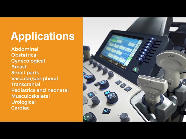
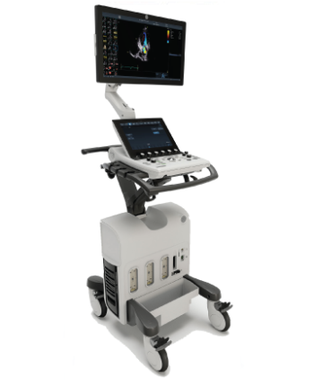
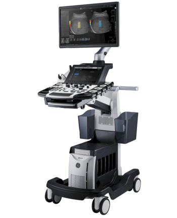
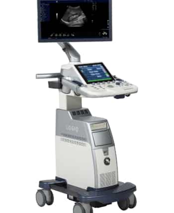
The GE Logiq S8 is a 4D multi-purpose ultrasound machine from GE’s “Signature” series of compact, mid-range ultrasounds. The Logiq S8 is designed for abdominal, vascular, cardiac, breast, small parts, obstetrics, gynecology, neonatal, urology, pediatrics, and transcranial applications. It is extremely light and compact, yet powerful enough to meet your ultrasound imaging needs, and looks visually similar to the Voluson S6, Voluson S8, and Logiq S7, which is its replacement. The Logiq S8’s powerful ultrasound system aims to get you the best images with minimal keystrokes. It minimizes workflows with its ergonomic design and convenience when scanning, so you can devote more time to patient care.
Read More