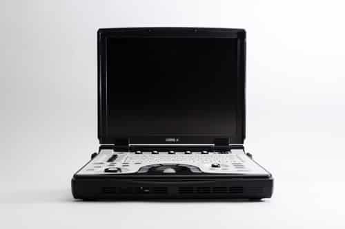
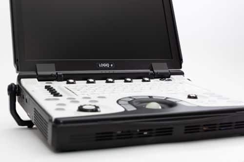
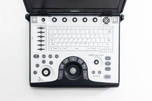
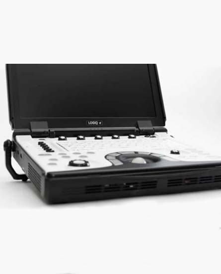
GE Logiq e BT12 Dimensions and Weight:
• Height: 70 mm (2.75 in) console only 100 mm (3.94 in) with handle
• Width: 340 mm (13.38 in)
• Depth: 346 mm (11.63 in) console only 372 mm (14.4 in) with handle
• Weight with Battery: approx. 5.2 kg (11.5 lb.)
GE Logiq e BT12 Specifications:
• Digital beamformer
• Displayed imaging depth: 0 – 30 cm (probe dependent)
• Minimum depth of field: 0 – 1 cm (probe dependent)
• Maximum depth of field: 0 – 30 cm (probe dependent)
• Continuous dynamic receive focus/continuous dynamic receive aperture
• Adjustable dynamic range upto 174dB
GE Logiq e BT12 Electrical power:
• Voltage: 100–240 VAC
• Frequency: 50/60 Hz
• Power: Max. 130 VA with Peripherals
GE Logiq e BT12 Review:
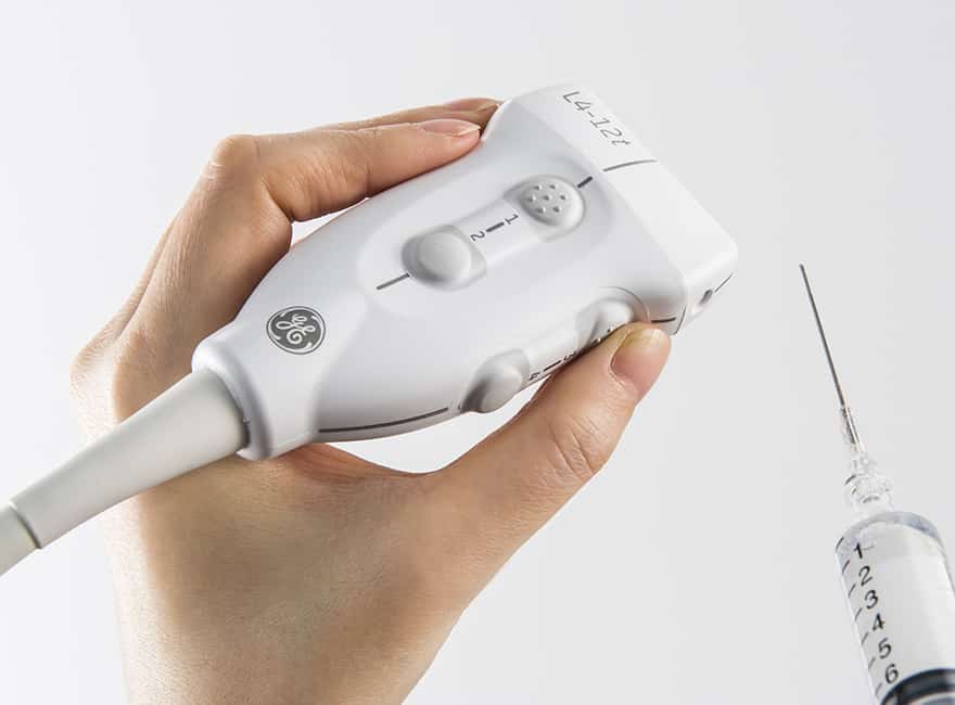
The GE Logiq e BT12 is designed for Point of Care and features GE’s new case color and design. Thanks to its hardware and software upgrades, it performs better than the GE Logiq e BT11 and also supports a high frequency hockey stick probe for orthopedics (musculoskeletal), rheumatology, and anesthesiology.
Physicians looking to own a portable ultrasound system that can handle general imaging, musculoskeletal, anesthesiology, interventional, emergency, and critical care applications, will find the GE Logiq e BT12 to be an excellent choice. It is also situated at a very attractive price point, and so for just for a couple of thousand dollars more than the GE Logiq e BT11, physicians can also consider the GE Logiq e BT12 as an affordable option.
GE Logiq e BT12 Probes/Transducers:
Convex Probe
4C-RS [2-5.5MHz]
Intra-Cavitary Probe
EC8C-RS [4-10MHz]
Micro-Convex Probe
8C-RS [4-10MHz]
Linear Probe
8L-RS [4-12MHz]
9L-RS [3.33-10MHz]
12L-RS [5-13MHz]
i12L-RS [4-10MHz]
L8-18i-RS [6.7-18MHz]
Sector
3Sc-RS [1.7-4MHz]
6S-RS [2.5-7MHz]
Speciality
i/T739L-RS [4-12MHz]
6Tc-RS [2-8MHz]
P2D [2MHz]
GE Logiq e BT12 Features:
• Laptop Style
• Integrated HDD (320 GB)
• CPU
– U7500 Core 2 Duo 1.06 G
• Lithium-Ion Battery Pack (standard)
– One hour scan time (mode dependent)
• Wired, wireless LAN Support
– For DICOM communication
– For Network Storage (Image store to PC without DICOM system)
• 1 probe port with micro-connector
• Rear handle
• High Resolution 15-inch Color LCD
• Over 1,000 frames or over 60 seconds CINE Memory (64 MB) depend on FOV, Scanning Lines etc.
• 320 GB Hard Drive
• External DVD R/W storage
• Loop storage—from live scanning and from memory
• Automatic Optimization
– Auto Tissue Optimization: ATO
– Auto Color Flow Optimization: ACO
– Auto Spectrum Optimization: ASO
– Auto Clarity Suite (Auto Focus and Auto Frequency)
• ACE* (Adaptive Color Enhancement)
• Raw Data Processing
• Patient Information Database
• Image Archive 97.5 GB on Hard Drive
• Full M&A Calculation Package with Real Time Auto Doppler Calculations
• Vascular Calcs
• Cardiac Calcs
• OB Calcs and Tables
• Fetal Trending
• Multi Gestational Calcs
• Musculoskeletal and Hip Dysplasia Calcs
• Gynecological Calcs
• Urological Calcs
• Renal Calcs
• Report Package
GE Logiq e BT12 Peripheral Options:
• Sony Digital UP-D711 Termal Printer
• Sony Fixture Kit for Digital UP-D711 Thermal Printer
• Sony Digital UP-D25 Color Thermal Printer
• Sony Digital UP-D897 BW Thermal Printer
• Mitsubishi P93W/E Thermal Printer
• Mitsubishi P95DW Thermal Printer
• 3 pedal Foot Switch (IPX8)
• 1 pedal Foot Switch (IPX8)
• NetGear USB Wireless Adapter WG111 V3 supporting the 802.11a/b/g formats, where available (option)
• Memory Stick (option)
• USB 2.0 Hub (option)
• USB HDD
• Barcode scanner (1D, 2D), HPP4600g
• Barcode scanner (1D), HHP3800g
GE Logiq e BT12 Supplies:
• Aquasonic ultrasound gel
• Sono ultrasound wipes
• Sony UPP-110HG thermal printing paper
• Sony UPC-21L color thermal printing pack
• Mitsubishi KP95HG thermal roll paper (new)
• Mitsubishi KP65HM-CE High density thermal paper
GE Logiq e BT12 ports:
• USB 2.0 x 2 ports
• Docking Cart
• Earphone Port
• Video Out(VGA)
• Ethernet
GE Logiq e BT12 image storage:
• On-board database of patient information from past exams
• Storage Formats:
– DICOM with ultrasound raw data
– Export JPEG and AVI formats
• Storage Devices:
– USB wireless LAN card
– USB HDD(1.5T Hard Drive)
– USB Memory Stick
– DVD R/W
– Hard Drive Image Storage: 320GB
– Network Storage support for Import, Export, DICOM Read, SaveAs, SaveAs
GE Logiq e BT12 Applications:
• Abdominal
• Cardiac
• Gynecology
• Intraoperative
• Musculoskeletal
• Obstetrical
• Nerve Block
• Pediatric and Neonatal
• Small Parts and Superficial
• Urological
• Vascular
GE Logiq e BT12 FAQs:
• Auto Optimization is a one-touch image optimization function that a user to optimize the image based on the actual B-mode image or Pulse wave Doppler data. The function works based on preset levels (Low, Medium, and High) and allows user to pick a preference for the contrast enhancement in the resulting image. Low does the least amount of contrast enhancement, high does the most. Auto Optimization is available in single or multi image, on live, frozen or CINE images (in B-Mode only), while in zoom, and in Spectral Doppler. Auto Optimization in PW Doppler Mode optimizes the spectral data. Auto adjusts the Velocity Scale (live imaging only), baseline shift, dynamic range, and invert (if preset). Upon deactivation, the spectrum is still optimized.
• CrossBeam (Spatial Compounding Imaging) obtains real time sonographic information from several different angles of insonation and combines them to produce a single image. CrossBeam helps reducing speckle artifacts, enhancing mass margin delineation, and improving anatomical details.
• Virtual Convex is available on linear and Sector probes. Virtual Convex provides a larger field of view in the far field. Virtual Convex is always active with sector probes.
• Raw Data is a software tool that enables image processing, quick data re-acquisition, and image analysis with same resolution and same frame rates of original images. Raw Data helps shorten exam duration, improves clinical work flow by post-processing, and reduces a time to put probe on a patient.
• LogiqView (Panoramic Imaging) enables a transducer to be moved along a larger organ, stitching multiple images together to form one long image with an extremely wide field of view.
• Auto IMT automates measurement of intima-media thickness of vessel. Auto IMT helps keep tracking atherosclerosis diseases from the early stage as it is developed.
• B Steer simply slants B-mode or Color Flow linear image left or right to get more information without moving the probe and enables enhanced needle visualization in real time for needle guidance procedures. The angle steer function only applies to linear probes.
• Anatomical M-Mode helps improve accuracy of arrhythmia assessments and cardiac measurements. It enables to provide M Mode images truly perpendicular to ventricular septum.
• eSmart Trainer Provides modules showing basic scanning techniques, with graphics of probe position, anatomy and example clinical image. Similar to Scan Assistant.
• Flow QA (Color and PDI Quantification) helps a user evaluate the amount of blood flow within a region of interest, to assist with diagnosis and monitoring.
• Patient Follow-up Tool when an user monitor the patient’s condition, therapy or progress over time – LOGIQ e automatically recalls all of the imaging parameters, comments and body patterns to be identical to the previous exam. It provides an alert if a user uses a different transducer than last time.